This guide explores the intricate world of portable X-ray fluorescence analyzers (XRF), delving into XRF training, which includes the certification process, radiation principles, exposure units, and safety measures.
Initiate your XRF operator certification journey now with Enviropass. Request your training today and secure your certification.
Specific safety protocols are typically essential for XRF handling, necessitating user training. XRF trainers must possess the requisite skills and knowledge. Two levels of XRF training are:
This is the first level of training, and operators with this certification are authorized to perform XRF operations. After the training, operators must pass an exam to demonstrate their understanding.
Level 2 operators, with additional responsibilities, can offer formal XRF training, which is beneficial for manufacturers and specialized training service companies. Operators at these levels must also pass the Level 2 exam, recognized by officials.

Natural Resources Canada (NRCan), a federal department overseeing certifications in non-destructive testing (NDT), manages the certification process for operators of XRF in Canada. Managed by the NRCan National NDT Certification Body (NDTCB), this process is part of the National NDT Personnel Certification Program, aligning with the Canadian General Standards Board (CGSB) standard CAN/CGSB-48.9712, which encompasses various NDT methods, including industrial radiography and ultrasonic testing.
In collaboration with Health Canada, NRCan has integrated the international standard ISO 20807:2004, tailoring modifications for X-ray operator certification. Health Canada, responsible for safety codes in federally regulated sectors, is crucial in shaping these modifications. Adhering to Safety Code 32, users of portable XRF devices must be NRCan XRF-certified operators. The certification program includes safety training and examinations, encompassing theory, usage, maintenance, storage, radiation protection, and safety considerations related to XRF.

Ordinarily, the training emphasizes safety for both the user and those in the vicinity due to the potential harm from emitted radiation. Here are the typical topics:
What are these radiations and their hazards? In XRF analysis, an XRF analyzer bombards the atoms on a metal surface with X-rays, leading to ionization. Ionization involves the dissociation of an atom or molecule into positively or negatively charged ions, essentially removing electrons from a neutral atom. X-rays and gamma rays can ionize matter, earning them the designation of ionizing radiation. This type of radiation can alter the chemical structure of living cells. Moreover, a potential risk is associated with X-rays backscattered from the analyzed sample.
XRF is one type of X-ray, which is fluorescent. It is typically emitted when materials are hit with X-rays. The nature of these materials influences the wavelength of XRF, revealing their composition.
Common XRF instruments include:
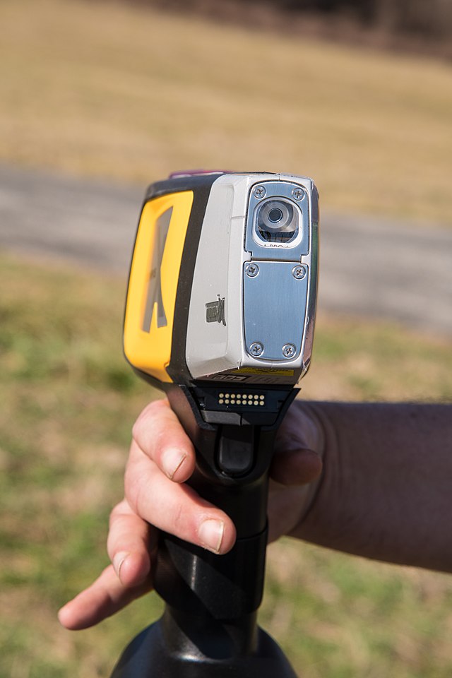
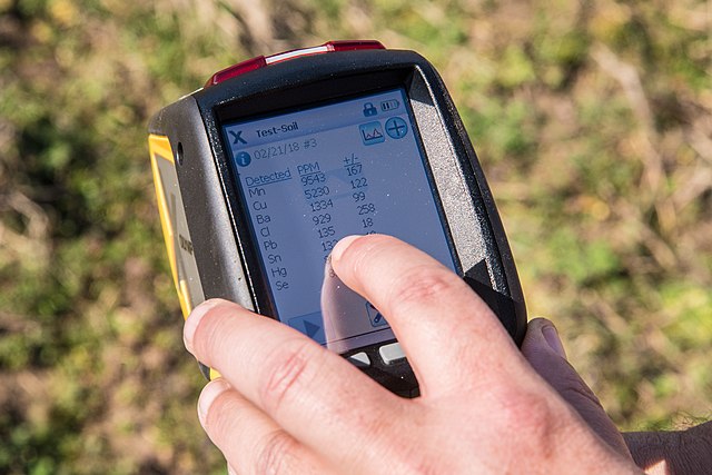
1- Portable XRF Analyzer: Portable and easy to use for on-site analysis in various industries for material identification, elemental analysis, and detection of heavy metals.
2-Benchtop XRF Spectrometers: Suitable for laboratory settings with accurate and precise results for various applications (part per million ppm level). For example, benchtop XRF spectrometers serve for hazardous substances determination. Therefore, these instruments help verify product environmental compliance of electronics, furniture, fabric, etc.
3- Wavelength Dispersive XRF (WDXRF) Spectrometers: Utilize crystal monochromators to separate and measure characteristic X-ray lines for precise elemental analysis.
4- XRF Coating Thickness Gauges: These gauges measure the thickness of coatings on various substrates, which is essential in industries like automotive and electronics manufacturing.
The roentgen unit is commonly used to measure the intensity of X-ray and gamma-ray radiation in the air. One roentgen (R) is the amount of X-rays or gamma rays needed to produce a certain amount of ionization in one cubic centimeter of dry air.
Unlike the roentgen for radiation in the air, the “dose unit” addresses the radiation absorbed by materials. It is a common term related to radiation exposure in the literature. In fact, it measures energy absorbed per unit mass when materials are exposed to ionizing radiation. This measure is crucial in assessing the impact of radiation on different substances. The two units for quantifying absorbed dose are:
Rad (Radiation Absorbed Dose): Rad provides a standardized measure for the amount of radiation energy absorbed by a specific substance. 1 Rad is equivalent to the absorption of 10 µJ of energy per gram of material.
Gray (International Standard Unit – SI): Gray is the globally recognized unit for absorbed dose, adhering to the International System of Units (SI). It simplifies international communication about radiation exposure and ensures consistency in measurements. 1 Gray equals 100 Rads.
Dose equivalence units were proposed based on the ability to cause biological damage. The Roentgen Equivalent Man (rem) measures the biological impact of ionizing radiation, considering both the absorbed dose and the specific type of radiation. It incorporates a weighting factor to quantify the potential harm, with one rem equivalent to one rad for X-rays and gamma rays. Rem is crucial in assessing health risks associated with exposure to ionizing radiation in radiological protection.
In summary: 1 Sievert (Sv) = 1 Gray = 100 rem = 100 Rad.
Humans can experience different levels of radiation exposure. Small amounts received over an extended period constitute a chronic dose. High radiation exposure can manifest various symptoms of radiation injury, including fatigue, vomiting, and hair loss. Large, short-term doses, known as acute doses, lead to physical and massive effects. In such cases, body cells lack sufficient time for repair, resulting in prolonged effects. Exposure exceeding 10 rem can lead to reduced blood count and hair loss. Recovery from acute doses can take up to a few months. Exposure beyond 100 rem causes significant changes in the body, known as radiation sickness. Exposure exceeding 500 rem, termed the Mean Lethal Dose (MLD), makes it impossible for the body to repair.
Generally, the body tolerates chronic doses better than acute ones, allowing more cell repair time. These effects may include genetic impacts on chromosomes, somatic effects leading to cancer and tumors, and, in some instances, heritable effects passed down to subsequent generations.
Several factors influence the biological damage resulting from radiation exposure. The greater the total dose and dose rate, the more pronounced the biological effects. Additionally, the area exposed and the type of cells impacted can affect the body differently. Developing embryos and children are at the highest risk of exposure, while middle-aged adults exhibit the most minor sensitivity.
We consistently receive radiation from various natural sources, like radon gas in homes, terrestrial and cosmic radiation, and internal elements in food and water. In fact, a person in North America receives on average 365 mrem of radiation annually. This includes natural sources and artificial sources. (United States Nuclear Regulatory Commission)
Depending on the age or other factors, humans can tolerate a certain degree of radiations. In fact, official agencies worldwide agree on acceptable limits. For example, the International Commission on Radiological Protection (ICRP) sets the maximum safe radiation exposure for the public and radiation workers per standard ICRP 2007 Radiation Dose Limits. Accordingly, the general public has an annual whole-body exposure limit of 100 mrem and 5 rem for the skin, excluding natural, consumer products, and medical exposure. Nevertheless, radiation workers have an extended limit of 2 rem for the whole body and 50 rem for the skin. The extent of body exposure influences the biological effects of a given dose, with larger exposed areas leading to greater effects.
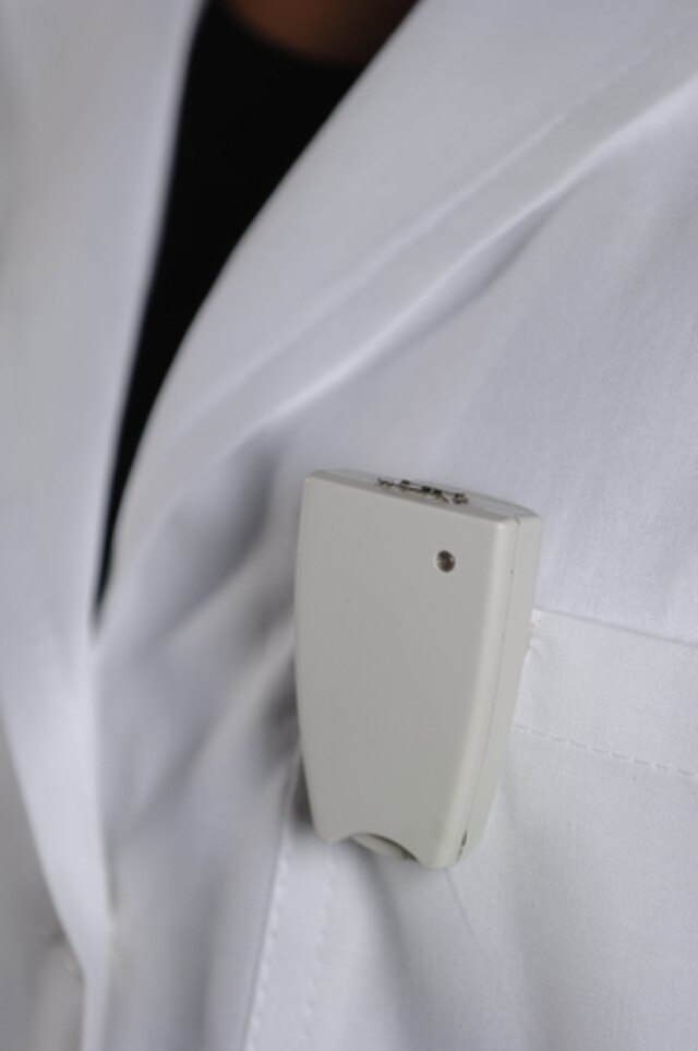
Special devices, commonly known as dosimeters, are designed to detect and record radiation exposure for personnel working with X-ray equipment. These devices come in various forms, such as:
To keep away our body from damages we have to consider the following ways to reduce the exposure from radiation:
The ALARA (As Low As Reasonably Achievable) concept is designed to prevent unnecessary exposure to employees and the public. It relies on the basic principles of time, distance, and shielding. Increasing the duration of exposure to radiation results in greater absorption. Conversely, maintaining a greater distance from the source of ionizing radiation leads to smaller doses since radiation loses energy as it travels through materials. Low X-ray radiation (< 50 keV – Kiloelectron volts) can easily penetrate and pass through samples.
The radiation intensity from a source is inversely proportional to the square of the distance from that source. For instance, doubling the distance reduces the intensity to ¼ of the original, and tripling the distance reduces it to 1/9. Further, using additional material as a barrier between a person and ionizing radiation results in receiving smaller doses. For example, lead and other heavy metals are common materials utilized in the walls of X-ray photography centers.
Minimize natural X-ray exposure by taking preventive measures against contributors like radon gas in homes and other sources by taking these precautions:
Consult local health authorities or radiation experts for specific concerns or areas with higher radiation levels. Adhere to safety guidelines from relevant health authorities and stay informed about potential risks linked to natural radiation sources.
In conclusion, certain safety practices necessitate careful consideration including:
The evolution of XRF devices from the 1950s marks a remarkable journey. Initially housed in heavily radioactive boxes, they have transformed into compact, safe designs for personnel use. This progression began with Wilhelm Roentgen’s accidental discovery of X-rays in 1895, earning him a Nobel Prize in Physics. Henry Moseley pioneered using X-rays for analysis in 1913, and by 1948, the first commercially available XRF emerged. In 1982, the initial handheld XRF became available, weighing 31 kg and requiring a trolley for movement. By 2001, significant optimization reduced its weight to 3 kg, enabling the measurement of magnesium as the lightest element.
Embark on your journey to becoming a certified XRF operator! Contact Enviropass today, and let our experienced team guide you through the certification process, ensuring your expertise in safe and efficient XRF operation.